Welcome to a deep dive into the fascinating world of cardiac muscle – the powerhouse behind your beating heart! In this blog post, we’ll unravel the secrets of what makes cardiac muscle unique, explore its intricate structure and function, and discover how it responds to various stimuli. So buckle up as we embark on a journey to uncover everything you need to know about the incredible cardiac muscle!
What is cardiac muscle?
Cardiac muscle, also known as myocardium, is a specialized type of muscle tissue found exclusively in the heart. It plays a crucial role in pumping blood throughout the body and maintaining circulation. Unlike skeletal muscles that are under voluntary control or smooth muscles found in organs like the intestines, cardiac muscle operates involuntarily to ensure continuous heart function.
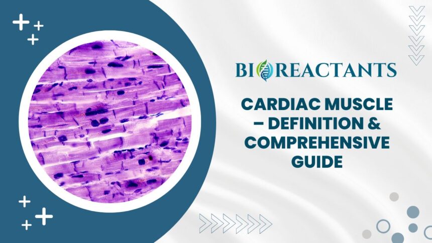
What sets cardiac muscle apart is its unique ability to contract rhythmically and efficiently without fatiguing. This constant contraction allows the heart to beat tirelessly, day after day, keeping us alive and well. Each cardiac muscle cell contains myofibrils packed with proteins responsible for generating force during contraction.
The interconnected network of cardiac muscle cells forms a syncytium that enables coordinated contractions across the entire heart. These cells communicate through specialized junctions called intercalated discs which facilitate rapid electrical impulses transmission essential for synchronous heartbeats.
How does cardiac muscle differ from skeletal and smooth muscle?
Cardiac muscle, also known as myocardium, is a specialized type of muscle found only in the heart. Unlike skeletal muscle, which is attached to bones and under voluntary control, cardiac muscle is involuntary and contracts rhythmically without conscious effort.
Another key difference lies in the structure of these muscles. While skeletal muscles are long fibers with multiple nuclei located at the periphery of each cell, cardiac muscles are shorter branched cells with a single nucleus centrally located.
Smooth muscle differs from both cardiac and skeletal muscles in terms of appearance and function. Smooth muscle cells lack striations seen in cardiac and skeletal muscles, giving them a smooth appearance under the microscope. Additionally, smooth muscle is found in organs like the intestines and blood vessels where it helps regulate movements such as peristalsis and vasoconstriction.
Each type of muscle serves a unique purpose within the body – whether it’s providing movement (skeletal), pumping blood (cardiac), or controlling involuntary functions (smooth).
What are the unique properties of cardiac muscle cells?
Cardiac muscle cells are distinct in their ability to contract rhythmically without fatigue, allowing the heart to continuously pump blood throughout the body. One unique property of cardiac muscle cells is their intercalated discs, specialized junctions that help synchronize contractions between neighboring cells.
These cells have a high density of mitochondria, providing them with ample energy for consistent contraction. Unlike skeletal muscles, cardiac muscle cells are branched and interconnected, forming a network that ensures coordinated pumping action.
Another key feature is their automaticity – they can generate electrical impulses independently of external stimuli. This intrinsic pacemaker activity allows the heart to maintain its own rhythm even in the absence of neural input.
These properties make cardiac muscle cells vital for sustaining life by enabling efficient circulation and oxygen delivery to all tissues.
How does the structure of cardiac muscle contribute to its function?
The structure of cardiac muscle is finely tuned to support its vital function in the heart. Unlike skeletal muscles, which are under voluntary control, cardiac muscles work involuntarily to maintain a steady heartbeat. The branching nature of cardiac muscle fibers allows for efficient transmission of electrical impulses throughout the heart.
These interconnected fibers form a syncytium that contracts as a single unit, ensuring coordinated pumping action. Additionally, the presence of intercalated discs between adjacent cells facilitates rapid communication and synchronization during contraction.
The abundance of mitochondria in cardiac muscle cells provides them with the energy needed to sustain constant activity without fatigue. Moreover, the striated appearance of cardiac muscle fibers reflects their organized arrangement for effective force generation.
The unique structural characteristics of cardiac muscle enable it to perform its crucial role in maintaining circulation and supporting overall cardiovascular health.
What role do intercalated discs play in cardiac muscle function?
Intercalated discs are like the secret sauce of cardiac muscle function, holding everything together in perfect harmony. These specialized junctions allow individual muscle cells to communicate and coordinate their contractions seamlessly.
Think of intercalated discs as the glue that keeps the heart pumping rhythmically and efficiently. They not only transmit electrical signals between cells but also ensure synchronized contraction so that the heart can effectively pump blood throughout the body.
By forming a strong connection between adjacent cardiac muscle cells, intercalated discs help maintain structural integrity and promote efficient communication within the heart tissue. This coordinated teamwork is essential for the heart to contract as a single unit, allowing it to beat in unison and propel blood forward with each rhythmic pulse.
Intercalated discs play a crucial role in ensuring that your heart functions like a well-oiled machine, keeping you alive and thriving every day without missing a beat or skipping a step.
How do cardiac muscle cells contract?
Cardiac muscle cells contract in a coordinated manner to facilitate the pumping action of the heart. Unlike skeletal muscles that require external stimulation, cardiac muscles can generate their own electrical impulses through specialized cells called pacemaker cells. These impulses travel through the heart and trigger muscle contraction.
The process begins with an electrical signal originating from the sinoatrial (SA) node, known as the heart’s natural pacemaker. This signal spreads across the atria, causing them to contract and push blood into the ventricles. The signal then reaches the atrioventricular (AV) node, which delays it slightly before passing it on to ensure proper timing.
From there, the impulse travels down special pathways in the heart called bundle branches and Purkinje fibers, rapidly stimulating all parts of both ventricles simultaneously. This synchronized contraction allows for efficient ejection of blood from both chambers and ensures an effective pump function crucial for maintaining circulation throughout your body.
By understanding how cardiac muscle cells contract harmoniously with each beat, we appreciate just how intricately designed our cardiovascular system truly is.
What is the significance of the myocardium in the heart?
The myocardium plays a crucial role in the heart’s function. It is the middle layer of the heart wall, composed mainly of cardiac muscle cells. These specialized cells are responsible for generating the force needed to pump blood throughout the body.
Within the myocardium, individual cardiac muscle cells work together in a synchronized manner to create a coordinated contraction and relaxation mechanism. This ensures that blood is efficiently circulated through the chambers of the heart and out into circulation.
The unique structure of the myocardium allows it to withstand constant stress and pressure without fatiguing easily. This durability is essential as the heart beats around 100,000 times per day on average.
Without a healthy myocardium, proper heart function would be compromised, leading to various cardiovascular issues. Therefore, maintaining optimal myocardial health is vital for overall well-being and longevity.
How do electrical impulses travel through cardiac muscle?
When it comes to understanding how electrical impulses travel through cardiac muscle, it’s like unraveling the intricate wiring of the heart’s communication system. These impulses originate from a cluster of cells called the sinoatrial (SA) node, often referred to as the heart’s natural pacemaker.
From the SA node, the electrical signals travel along specialized pathways within the myocardium, known as Purkinje fibers and intercalated discs. These structures ensure rapid and coordinated transmission of impulses throughout the heart muscle.
As these electrical signals move through cardiac muscle cells, they trigger contractions that pump blood efficiently throughout our bodies. It’s this synchronized dance of electrical activity and muscular response that keeps our hearts beating steadily day in and day out.
Understanding how these impulses flow through cardiac muscle sheds light on just how remarkable our hearts truly are – functioning tirelessly to keep us alive and well with every beat.
What is the role of the sinoatrial (SA) node in cardiac muscle activity?
The sinoatrial (SA) node, often referred to as the heart’s natural pacemaker, plays a crucial role in regulating cardiac muscle activity. Located in the right atrium of the heart, this specialized cluster of cells generates electrical impulses that initiate each heartbeat. These signals travel through the atria, causing them to contract and push blood into the ventricles.
As the SA node fires its electrical impulses, it sets the pace for the heart’s rhythm. This rhythmic activity is essential for maintaining proper blood flow throughout the body. By coordinating the timing of contractions between different chambers of the heart, the SA node ensures efficient pumping action.
In addition to setting the heart rate, the SA node also responds to signals from other parts of the autonomic nervous system. This allows for adjustments in heart rate based on factors like stress levels or physical exertion. The intricate interplay between these regulatory mechanisms helps maintain cardiovascular homeostasis and adaptability.
Understanding how this small but mighty structure functions within our hearts sheds light on its vital contribution to our overall health and well-being.
How does the autonomic nervous system influence cardiac muscle function?
The autonomic nervous system plays a crucial role in regulating the function of cardiac muscle. It’s like a silent conductor orchestrating the rhythm of your heart behind the scenes.
Through its sympathetic and parasympathetic branches, it can speed up or slow down your heartbeat depending on the body’s needs. Think of it as a dance between fight-or-flight responses and rest-and-digest modes.
When you’re under stress or exerting yourself during exercise, the sympathetic nervous system kicks in to increase heart rate and contractility, ensuring that your muscles receive enough oxygen-rich blood.
Conversely, during times of relaxation or sleep, the parasympathetic system takes over to decrease heart rate and promote calmness. It’s like a gentle lullaby for your heart.
This intricate interplay between the autonomic nervous system and cardiac muscle keeps your ticker ticking in perfect harmony without you even having to think about it.
What are the effects of exercise on cardiac muscle?
Regular exercise has a profound impact on cardiac muscle. When you engage in physical activity, your heart works harder to pump blood efficiently throughout the body. This increased demand leads to adaptations in the cardiac muscle, making it stronger and more efficient over time.
Through regular exercise, cardiac muscle undergoes beneficial changes such as increased stroke volume and improved oxygen delivery capacity. These enhancements allow the heart to beat more effectively during both rest and activity, promoting overall cardiovascular health.
Additionally, exercise can help reduce the risk of developing heart disease by strengthening the heart muscle and improving its ability to respond to stressors. It also plays a role in maintaining healthy blood pressure levels and cholesterol profiles, further supporting optimal cardiac function.
Incorporating regular aerobic activities like running, cycling, or swimming into your routine can significantly benefit your cardiac muscle and overall heart health. Remember that consistency is key when it comes to reaping the benefits of exercise on your cardiovascular system.
How does cardiac muscle adapt to increased workload or stress?
Cardiac muscle is a remarkable tissue that can adapt to increased workload or stress in impressive ways. When faced with heightened demand, such as during exercise or in conditions like hypertension, cardiac muscle undergoes changes to meet the body’s needs. One way it adapts is by increasing the size of individual cardiac muscle cells through a process known as hypertrophy.
This enlargement allows the heart to pump more effectively and efficiently against resistance. Additionally, cardiac muscle can enhance its contractile strength and optimize energy production pathways to sustain prolonged periods of heightened activity. These adaptations help ensure that the heart continues to function optimally even under duress.
Moreover, increased workload prompts physiological responses that promote better oxygen delivery and utilization within the cardiac muscle cells. This improved efficiency enables the heart to cope with greater demands without compromising its performance or health.
What are common diseases and conditions that affect cardiac muscle?
Cardiac muscle, despite its remarkable strength and endurance, is not invincible. There are several diseases and conditions that can impact the health of this vital muscle in our bodies. One common condition is coronary artery disease, where plaque buildup restricts blood flow to the heart, leading to potential damage or even a heart attack.
Another prevalent issue is arrhythmias, which are irregular heart rhythms that can disrupt the efficient pumping action of the heart. Heart failure is also a significant concern, where the heart struggles to pump enough blood to meet the body’s needs. This can result from various factors such as high blood pressure or previous heart attacks.
Furthermore, cardiomyopathy refers to diseases affecting the myocardium itself, weakening the cardiac muscle over time. Additionally, valvular heart disease involves problems with one or more of the heart valves interfering with proper blood flow through the chambers.
It’s crucial to be aware of these conditions and seek medical attention if experiencing symptoms like chest pain, shortness of breath or palpitations. Regular check-ups and a healthy lifestyle play essential roles in maintaining cardiac muscle health for overall well-being.
How is cardiac muscle damage diagnosed and treated?
When it comes to diagnosing cardiac muscle damage, healthcare providers often utilize various tests and imaging techniques. These may include electrocardiograms (ECGs), echocardiograms, cardiac MRI scans, and coronary angiography to assess the extent of damage.
Treatment for cardiac muscle damage typically involves a combination of medications, lifestyle changes, and in some cases, surgical interventions. Medications such as beta-blockers, ACE inhibitors, or blood thinners may be prescribed to manage symptoms and prevent further complications.
In more severe cases of cardiac muscle damage like heart attacks or advanced heart failure, procedures such as angioplasty with stent placement or even heart transplant surgery might be necessary. Cardiac rehabilitation programs can also play a crucial role in helping patients recover from cardiac muscle damage by focusing on exercise training and education about heart-healthy habits.
What are the latest advancements in cardiac muscle research and therapy?
Exciting developments in cardiac muscle research and therapy are continuously emerging, paving the way for innovative treatments and improved patient outcomes. Researchers are exploring cutting-edge techniques such as gene editing to target specific cardiac conditions at a molecular level.
Advancements in stem cell therapy offer promising possibilities for regenerating damaged heart tissue, potentially revolutionizing treatment options for heart failure patients. Additionally, the use of artificial intelligence and machine learning algorithms is enhancing diagnostic accuracy by analyzing vast amounts of cardiovascular data.
Novel drug therapies tailored to individual genetic profiles show great potential in optimizing treatment efficacy while minimizing side effects. Furthermore, advancements in medical devices like implantable cardioverter-defibrillators are transforming how we manage arrhythmias and sudden cardiac events.
The integration of telemedicine services into cardiac care allows for remote monitoring and timely interventions, improving access to specialized care regardless of geographical location. Stay tuned as researchers continue to push boundaries in understanding and treating cardiac muscle disorders with unparalleled innovation!
Conclusion
The cardiac muscle is a remarkable and vital component of our bodies, ensuring that our hearts beat effectively to pump blood throughout our system. Understanding the intricacies of how cardiac muscle functions can help us appreciate its importance in maintaining overall health. With ongoing research and advancements in treatment options, we continue to deepen our knowledge of cardiac muscle physiology and find innovative ways to address related conditions. By prioritizing heart health through lifestyle choices like exercise and proper nutrition, we can support the strength and resilience of our cardiac muscle for a healthier future.


